IAV
Trial Size

IRAK Antibody

CATALOG NUMBER: 1007

Clonality:
Polyclonal
Tested Applications:
ELISA, ICC, IF, IP, WB
Host Species:
Rabbit
Species Reactivity:
Human, Mouse, Rat
Conjugate:
Unconjugated
Specifications
Host Species:
Rabbit
Species Reactivity:
Human, Mouse, Rat
Homology:
Predicted species reactivity based on immunogen sequence: Bovine: (85%)
Immunogen:
Anti-IRAK antibody (1007) was raised against a peptide corresponding to 13 amino acids near the carboxy terminus of human IRAK.
The immunogen is located within the last 50 amino acids of IRAK.
Conjugate:
Unconjugated
Tested Applications:
ELISA, ICC, IF, IP, WB
Application Note:
WB: 1-4 μg/mL; IF: 20 μg/mL; ICC: 10 μg/mL.
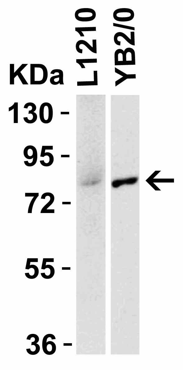
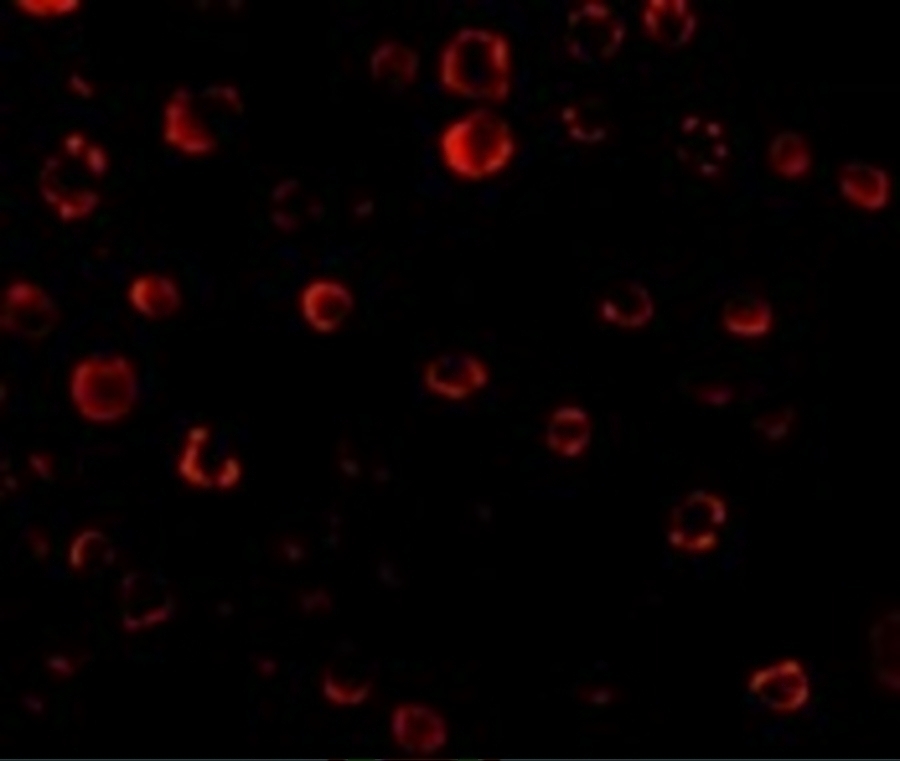
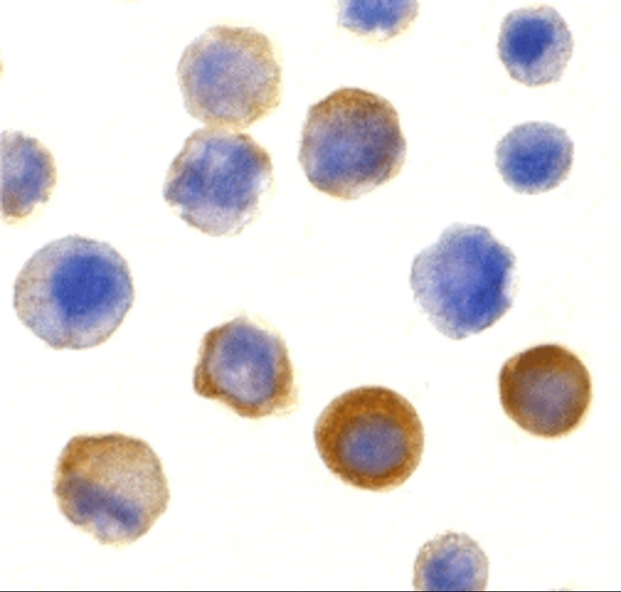
Antibody validated: Western Blot in human, mouse and rat samples; Immunofluorescence and Immunocytochemistry in human samples. All other applications and species not yet tested.
Specificity:
At least four isoforms of IRAK are known to exist; this antibody will detect all four isoforms. IRAK antibody is predicted to not cross-react with other members of the IRAK protein family.
Positive Control 1:
Cat. No. 1201 - HeLa Cell Lysate
Positive Control 2:
Cat. No. 17-001 - HeLa Cell Slide
Positive Control 3:
Cat. No. 1220 - SK-N-SH Cell Lysate
Predicted Molecular Weight:
Predicted: 77kD
Observed: 77 kD
Validation:

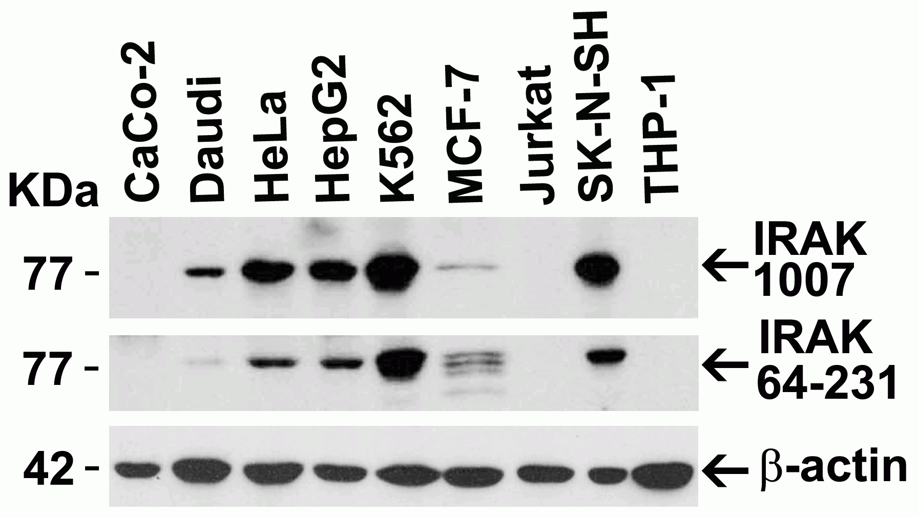
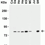
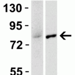
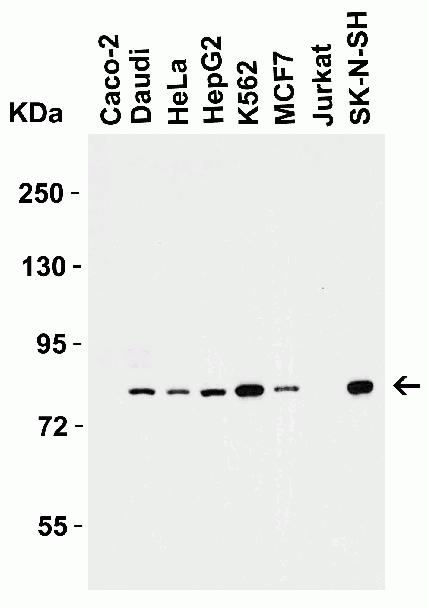
Independent Antibody Validation in Cell lines (Figure 2) shows similar IRAK expression profile in human cell lines detected by two independent anti-IRAK antibodies that recognize different epitopes, 1007 against C-terminus domain and 64-231 against another region of C-terminus domain.  IRAK proteins are detected in the most tested cell lines at different expression levels by the two independent antibodies.

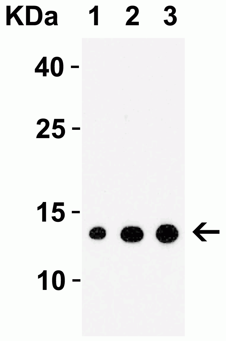
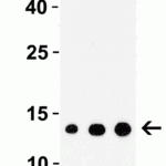
Recombinant Protein Test (Figure 3): Anti-IRAK antibodies (1007) detected human IRAK recombinant protein at different concentrations.

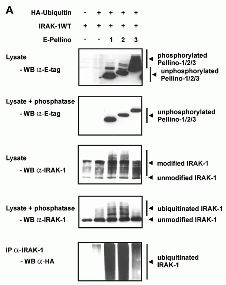
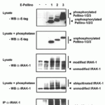
Immunoprecipitation validation (Figure 6): IRAK1 protein and pellino protein were immunoprecipitated by anti-IRAK antibodies (1007) in HEK293T cells with co-expression of Pellino proteins and IRAK-1.

Overexpression validation (Figure 7): IRAK1 overexpression in 293T cells was detected by anit-IRAK antibodies (1007).

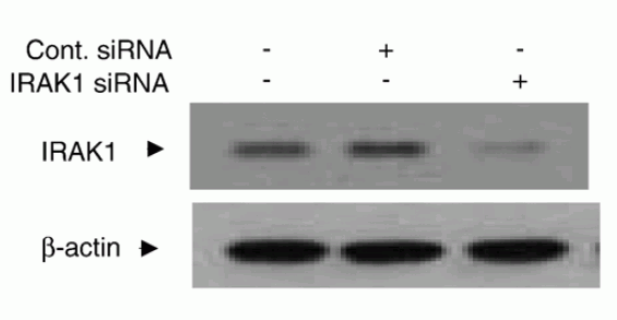
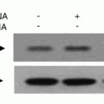
KD validation (Figure 8): Anti-IRAK antibody (1007) specificity was further verified by IRAK specific knockdown. IRAK signal in human chondrocytes transfected with IRAK siRNAs was disrupted in comparison with that in cells transfected with control siRNAs.

Isoforms:
Human IRAK has 4 isoforms, including isoform 1 (712aa, 77kD), isoform 2 (682aa, 73kD), isoform 3 (693aa, 75kD), and isoform 4 (633aa, 68kD). Mouse IRAK has 2 isoforms, including isoform 1 (710aa, 77kD) and isoform 2 (712aa, 78kD). Rat IRAK also has one isoform (710aa, 78kD). 1007 can detect all human isoforms, mouse and rat IRAK.
Purification:
IRAK Antibody is affinity chromatography purified via peptide column.
Clonality:
Polyclonal
Isotype:
IgG
physical-state:
Liquid
Buffer:
IRAK Antibody is supplied in PBS containing 0.02% sodium azide.
Concentration:
1 mg/mL
Storage Conditions:
IRAK antibody can be stored at 4°C for three months and -20°C, stable for up to one year. As with all antibodies care should be taken to avoid repeated freeze thaw cycles. Antibodies should not be exposed to prolonged high temperatures.
Ncbi Official Symbol:
IRAK1
Additional Names:
IRAK Antibody: IRAK, pelle, IRAK, Interleukin-1 receptor-associated kinase 1, IRAK-1
Protein Accession Number:
P51617
Protein Gi Number:
8928535
Ncbi Gene Id Number:
3654
User Note:
Optimal dilutions for each application to be determined by the researcher.
Background:
IRAK Antibody: Nuclear factor kappa B (NF-κB) is a ubiquitous transcription factor and an essential mediator of gene expression during activation of immune and inflammatory responses. NF-κB mediates the expression of a great variety of genes in response to extracellular stimuli including IL-1, TNFα and LPS. A serine/threonine protein kinase associated with IL-1 receptor (IRAK) and its homologue mouse pelle-like protein kinase (mPLK) were identified recently. IRAK is associated with the IL-1 receptor subunits IL-1RI and IL-1RAcP after IL-1 binding and serves as a signaling molecule to mediate IL-1 response. IRAK mediates a signaling cascade leading to NF-κB activation by members in IL-1 family including IL-1 and a novel cytokine IL-18 (also termed IGIF).
Background Reference 1:
Cao et al. Science 1996;271:1128-31.
Background Reference 2:
Trofimova et al. J Bio Chem 1996; 271: 17609-1.
Background Reference 3:
Huang et al. Proc Natl Acad Sci USA 1997;94:12829-12832.
Background Reference 4:
Robinson et al. Immunity 1997;7:571-581.
Citation 1:
Schauvliege et al. Pellino proteins are more than scaffold proteins in TLR/IL-1R signalling: a role as novel RING E3-ubiquitin-ligases. FEBS Lett. 2006;580(19):4697-702. PMID: 16884718
Citation 2:
Ahmad et al. MyD88, IRAK1 and TRAF6 knockdown in human chondrocytes inhibits interleukin-1-induced matrix metalloproteinase-13 gene expression and promoter activity by impairing MAP kinase activation. Cell Signal. 2007 ;19(12):2549-57. PMID: 17905570
Citation 3:
Ahmad et al. Elevated expression of the toll like receptors 2 and 4 in obese individuals: its significance for obesity-induced inflammation. J Inflamm (Lond). 2012;9(1):48. PMID: 23191980

FOR RESEARCH USE ONLY

For additional information, visit ProSci’s Terms & Conditions Page.

Disclaimer:
Optimal dilutions/concentrations should be determined by the end user. The information provided is a guideline for product use. This product is for research use only.

CATALOG NUMBER:

1007

List Size:
0.02 mg, 0.1 mg

List Price:

Price range: $99.00 through $445.00

Shipping Info
Customize your order
Custom Antibody Services

New & Featured Products

CATALOG NUMBER: PM-7369-HRP

CATALOG NUMBER: PM-7369-biotin

CATALOG NUMBER: PM-7369