IAV
KD
Most Cited
Most Images
Trial Size

PD-L1 Antibody

CATALOG NUMBER: 4059

Clonality:
Polyclonal
Tested Applications:
ELISA, Flow, IF, IHC-P, WB
Host Species:
Rabbit
Species Reactivity:
Human, Mouse, Rat
Conjugate:
Unconjugated
Specifications
Host Species:
Rabbit
Species Reactivity:
Human, Mouse, Rat
Homology:
Predicted species reactivity based on immunogen sequence: Rat: (77%), Mouse: (71%)
Immunogen:
Anti-PD-L1 antibody (4059) was raised against a peptide corresponding to 17 amino acids near the center of human PD-L1 isoform 1.
The immunogen is located within amino acids 60 - 110 of PD-L1.
Conjugate:
Unconjugated
Tested Applications:
ELISA, Flow, IF, IHC-P, WB
Application Note:
WB: 1-2 μg/mL; IHC-P: 2.5-5 μg/mL; IF: 20 μg/mL; Flow Cyt: 0.5 μg/mL.
Antibody validated: Western Blot in human and mouse samples; Immunohistochemistry in human and rat samples; Immunofluorescence in human and rat samples and Flow Cytometry in mouse samples. All other applications and species not yet tested.
Specificity:
PD-L1 antibody has no cross-reactivity to PD-L2.
Positive Control 1:
Cat. No. 1201 - HeLa Cell Lysate
Positive Control 2:
Cat. No. 1224 - Daudi Cell Lysate
Positive Control 3:
Cat. No. 1204 - K562 Cell Lysate
Positive Control 4:
Cat. No. 10-501 - Human Heart Tissue Slide
Positive Control 5:
Cat. No. 1301 - Human Heart Tissue Lysate
Predicted Molecular Weight:
Predicted: 33 kDa
Observed: 37 kDa
Validation:

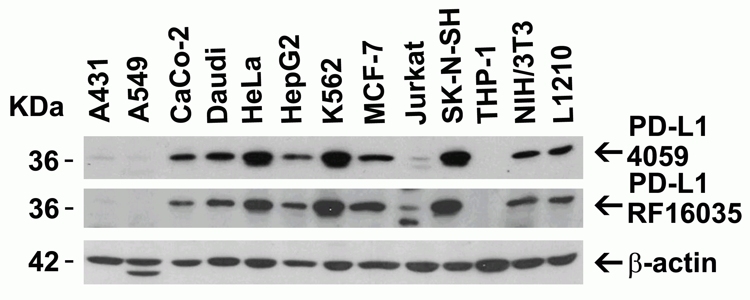
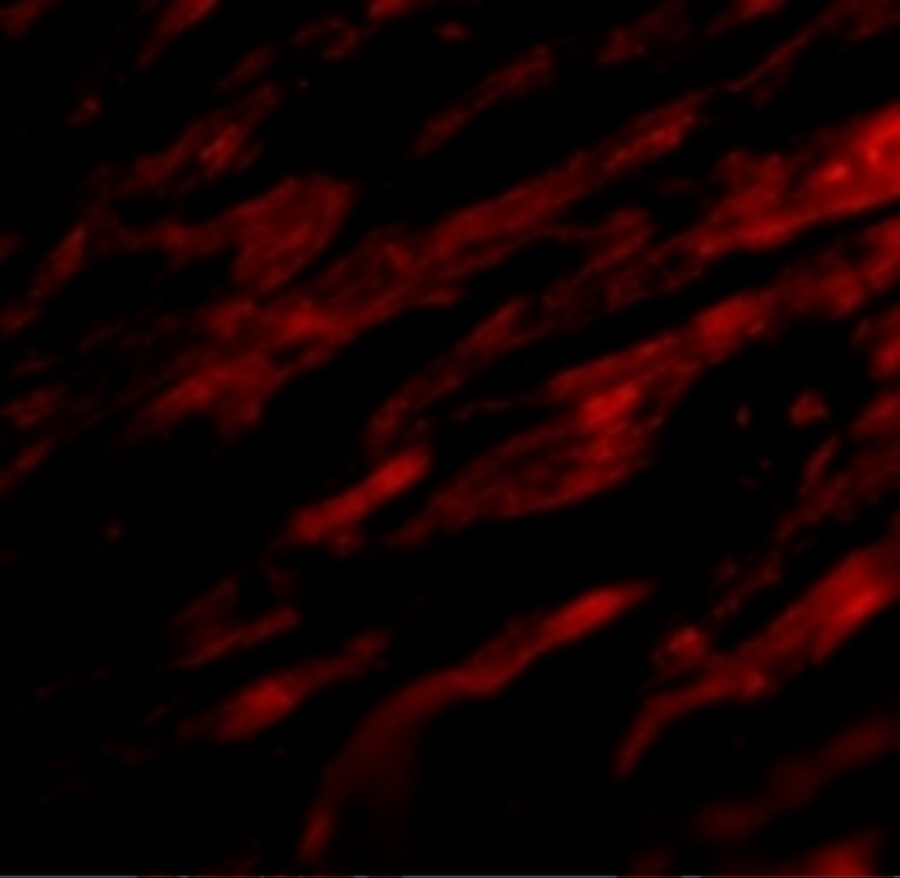
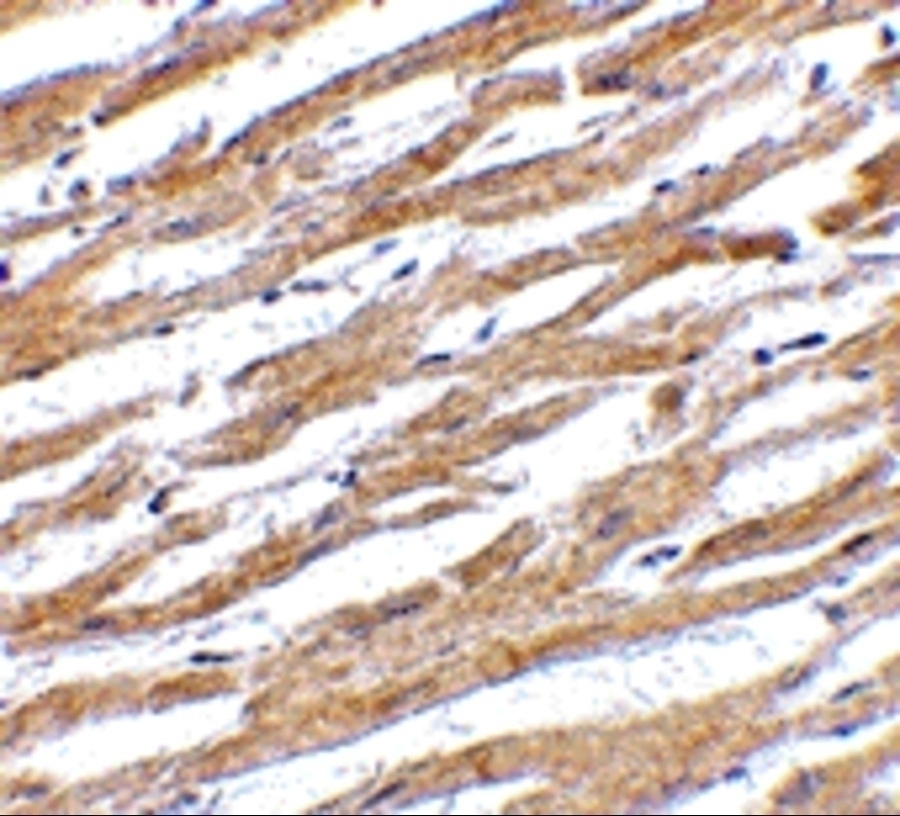
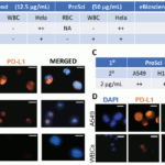
Independent Antibody Validation (Figure 2) shows similar PD-L1 expression profile in both human and mouse cell lines detected by two independent anti-PD-L1 antibodies that recognize different epitopes, 4059 against the center of human PD-L1 and RF16035 against the extracellular domain.  PD-L1 proteins are detected in most of the cell lines, but not in A549 and THP-1 cells by the two independent antibodies. 

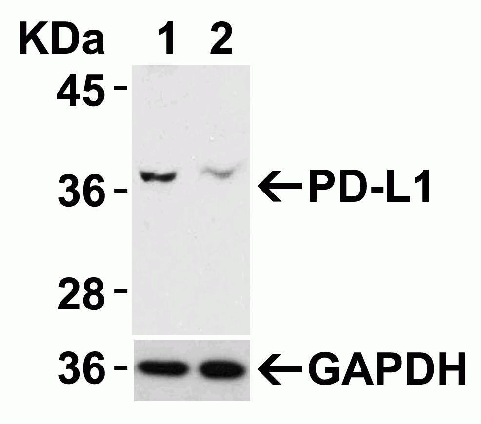
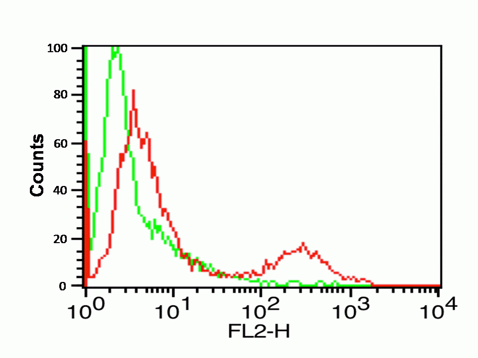
siRNA Knockdown Validation (Figure 3): Anti-PD-L1 antibody (4059) specificity was further verified by PD-L1 specific siRNA knockdown. PD-L1 signal in HeLa cells transfected with PD-L1 siRNAs was much weaker in comparison with that in HeLa cells transfected with control siRNAs.

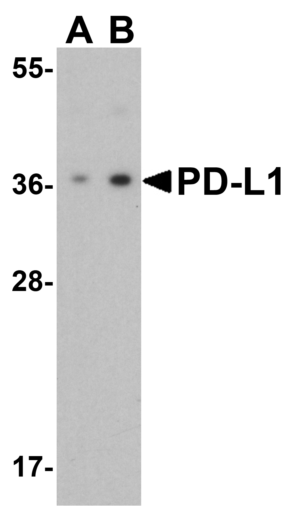
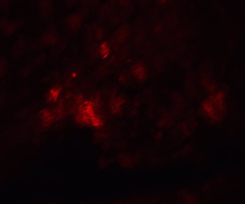
Overexpression Validation (Figure 4): Anti-PD-L1 antibodies (4059) can detect the overexpression of PD-L1 protein in 293 cells transfected with PD-L1.

Isoforms:
Human PD-L1 has 3 isoforms, including isoform 1 (290aa, 33.3kD), isoform 2 (176aa, 20.2kD), and isoform 3 (178aa, 20.5kD). This antibody detects human isoform 1&3, mouse and rat PD-L1 (290aa, 33kD for both of them).
Purification:
PD-L1 Antibody is affinity chromatography purified via peptide column.
Clonality:
Polyclonal
Isotype:
IgG
physical-state:
Liquid
Buffer:
PD-L1 Antibody is supplied in PBS containing 0.02% sodium azide.
Concentration:
1 mg/mL
Storage Conditions:
PD-L1 antibody can be stored at 4°C for three months and -20°C, stable for up to one year. As with all antibodies care should be taken to avoid repeated freeze thaw cycles. Antibodies should not be exposed to prolonged high temperatures.
Ncbi Official Symbol:
CD274
Additional Names:
PD-L1 Antibody: B7-H, B7H1, PDL1, PD-L1, PDCD1L1, PDCD1LG1, Programmed cell death 1 ligand 1, B7 homolog 1
Protein Accession Number:
NP_054862
Protein Gi Number:
7661534
Ncbi Gene Id Number:
29126
User Note:
Optimal dilutions for each application to be determined by the researcher.
Background:
PD-L1 plays a critical role in induction and maintenance of immune tolerance to self. As a ligand for the inhibitory receptor PDCD1/CD279, PD-L1 modulates the activation threshold of T-cells and limits T-cell effector response (1). The PDCD1/CD279-mediated inhibitory pathway is exploited by tumors to attenuate anti-tumor immunity and facilitate tumor survival (2,3). Through a yet unknown activating receptor, it may costimulate T-cell subsets that predominantly produce interleukin-10 (IL10) (4).
Background Reference 1:
Freeman et al. Exp. Med. 2000; 192:1027-34.
Background Reference 2:
Burr et al. Nature 2017; 549:101-5.
Background Reference 3:
Mezzadra et al. Nature 2017; 549:106-10.
Background Reference 4:
Dong et al. Nat. Med. 1999 5:1365-9.
Citation 1:
Dhar et al. Evaluation of PD-L1 expression on vortex-isolated circulating tumor cells in metastatic lung cancer. Sci Rep. 2018;8(1):2592. PMID: 29416054
Citation 2:
Gadiot et al. Overall survival and PD-L1 expression in metastasized malignant melanoma. Cancer.2011;117(10):2192-201. PMID: 21523733
Citation 3:
Angell et al. BRAF V600E in papillary thyroid carcinoma is associated with increased programmed death ligand 1 expression and suppressive immune cell infiltration. Thyroid. 2014; 24(9):1385-93PMID: 24955518
Citation 4:
Ilie et al. Comparative study of the PD-L1 status between surgically resected specimens and matched biopsies of NSCLC patients reveal major discordances: a potential issue for anti-PD-L1 therapeutic strategies. Ann Oncol. 2016;27(1):147-53.PMID: 26483045
Citation 5:
Heymann et al. PD-L1 expression in non-small cell lung carcinoma: Comparison among cytology, small biopsy, and surgical resection specimens. Cancer Cytopathol. 2017;125(12):896-907. PMID: 29024471
Citation 6:
Hamanishi et al. Safety and Antitumor Activity of Anti-PD-1 Antibody, Nivolumab, in Patients With Platinum-Resistant Ovarian Cancer. J Clin Oncol. 2015; 33(34):4015-22. PMID: 26351349
Citation 7:
Taube et al. Colocalization of inflammatory response with B7-h1 expression in human melanocytic lesions supports an adaptive resistance mechanism of immune escape. Sci Transl Med. 2012;4(127):127ra37. PMID: 22461641
Citation 8:
Munari et al. PD-L1 Expression Heterogeneity in Non-Small Cell Lung Cancer: Defining Criteria for Harmonization between Biopsy Specimens and Whole Sections. J Thorac Oncol. 2018;13(8): 1113-1120. PMID: 29704674
Citation 9:
Li et al. Comparison of 22C3 PD-L1 Expression between Surgically Resected Specimens and Paired Tissue Microarrays in Non-Small Cell Lung Cancer. J Thorac Oncol. 2017;12(10):1536-1543. PMID: 28751245
Citation 10:
Shen et al. Programmed cell death ligand 1 expression in osteosarcoma. Cancer Immunol Res. 2014;2(7):690-698. PMID: 24866169
Citation 11:
Mimura et al. PD-L1 expression is mainly regulated by interferon gamma associated with JAK-STAT pathway in gastric cancer. Cancer Sci. 2018 ;109(1):43-53.PMID: 29034543
Citation 12:
Habib et al. PDL-1 Blockade Prevents T Cell Exhaustion, Inhibits Autophagy, and Promotes Clearance of Leishmania donovani. Infect Immun. 2018;86(6). PMID: 29610255
Citation 13:
Gonin et al. Expression of IL-27 by tumor cells in invasive cutaneous and metastatic melanomas. PLoS One. 2013;8(10):e75694. PMID: 24130734
Citation 14:
Gagné et al. Comprehensive Assessment of PD-L1 Staining Heterogeneity in Pulmonary Adenocarcinomas Using Tissue Microarrays: Impact of the Architecture Pattern and the Number of Cores. Am J Surg Pathol. 2018;42(5):687-694.PMID: 29309297
Citation 15:
Teixidó et al. Assays for predicting and monitoring responses to lung cancer immunotherapy. Cancer Biol Med. 2015;12(2):87-95. PMID: 26175924
Citation 16:
Ohue et al. Survival of Lung Adenocarcinoma Patients Predicted from Expression of PD-L1, Galectin-9, and XAGE1 (GAGED2a) on Tumor Cells and Tumor-Infiltrating T Cells. Cancer Immunol Res. 2016;4(12):1049-1060. PMID: 27799141
Citation 17:
Kao et al. Assessing the Effects of Concurrent versus Sequential Cisplatin/Radiotherapy on Immune Status in Lung Tumor-Bearing C57BL/6 Mice. Cancer Immunol Res. 2015;3(7):741-50. PMID: 25672395
Citation 18:
Fujimura et al. Potential use of bisphosphonates in invasive extramammary Paget's disease: an immunohistochemical Investigation. Clin Dev Immunol. 2013 ;2013:164982. PMID: 23606867
Citation 19:
Sakata et al. Comparison of Programmed Death Ligand-1 Immunohistochemical Staining Between Endobronchial Ultrasound Transbronchial Needle Aspiration and Resected Lung Cancer Specimens. Chest. 2018;154(4):827-837. PMID: 30059678
Citation 20:
Kitazono et al. Reliability of Small Biopsy Samples Compared With Resected Specimens for the Determination of Programmed Death-Ligand 1 Expression in Non--Small-Cell Lung Cancer. Clin Lung Cancer. 2015 ;16(5):385-90.PMID: 25937270

FOR RESEARCH USE ONLY

For additional information, visit ProSci’s Terms & Conditions Page.

Disclaimer:
Optimal dilutions/concentrations should be determined by the end user. The information provided is a guideline for product use. This product is for research use only.

CATALOG NUMBER:

4059

List Size:
0.02 mg, 0.1 mg

List Price:

Price range: $99.00 through $445.00

Shipping Info
Customize your order
Custom Antibody Services

New & Featured Products

CATALOG NUMBER: PM-7369-HRP

CATALOG NUMBER: PM-7369-biotin

CATALOG NUMBER: PM-7369