Western blot analysis of PD-L2 in overexpressing HEK293 cells PD-L2 antibody at 0.5 and 1 μg/ml
Trial Size
PDL2 Antibody [7C1]
CATALOG NUMBER: RF16024
Clonality:
Monoclonal
Tested Applications:
ELISA, Flow, ICC, IF, IHC-P, WB
Host Species:
Mouse
Species Reactivity:
Human
Conjugate:
Unconjugated
Specifications
Host Species:
Mouse
Species Reactivity:
Human
Immunogen:
PD-L2 antibody was raised against the extracellular domain of human PD-L2.
Conjugate:
Unconjugated
Tested Applications:
ELISA, Flow, ICC, IF, IHC-P, WB
Application Note:
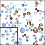
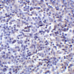
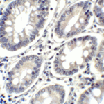
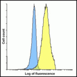
PD-L2 antibody can be used for detection of PD-L2 by Western blot at 0.5 - 1 μg/mL. Antibody can also be used for immunohistochemistry starting at 2 μg/mL. For immunofluorescence start at 20 μg/mL. Antibody validated: Western Blot in human samples; Immunohistochemistry in human samples; Immunocytochemistry in human samples; Immunofluorescence in human samples and Flow Cytometry in mouse samples. All other applications and species not yet tested.
Positive Control 1:
Cat. No. 1207 - Raji Cell Lysate
Positive Control 2:
Cat. No. 1315 - Human Tonsil Tissue
Predicted Molecular Weight:
Predicted: 30 kDa Observed: 38 kDa
Advanced Validation
Properties
Purification:
PD-L2 Antibody is supplied as protein A purified IgG2b.
Clonality:
Monoclonal
Isotype:
IgG2b
physical-state:
Liquid
Buffer:
PD-L2 Antibody is supplied in PBS containing 0.02% sodium azide and 50% glycerol.
Concentration:
1 mg/mL
Storage Conditions:
PD-L2 antibody can be stored at 4°C for three months and -20°C, stable for up to one year. As with all antibodies care should be taken to avoid repeated freeze thaw cycles. Antibodies should not be exposed to prolonged high temperatures.
Optimal dilutions for each application to be determined by the researcher.
Background
Background:
PD-L2 Antibody: Cell-mediated immune responses are initiated by T lymphocytes that are themselves stimulated by co gnate peptides bound to MHC molecules on antigen-presenting cells (APC). T-cell activation is generally self-limited as activated T cells express receptors such as PD-1 (also known as PDCD-1) that mediate inhibitory signals from the APC. PD-1 can bind two different but related ligands, PD-L1 and PD-L2, both of which are thought act as a negative regulator of T cell activation. However, it has been suggested that PD-L2 can act to stimulate an immunogenic response through and alternative receptor from PD-1.
Background Reference 1:
Holling TM, Schooten E, and van Den Elsing PJ. Function and regulation of MHC class II molecules in T-lymphocytes: of mice and men. Hum. Immunol. 2004; 65:282-90.
Background Reference 2:
Ishida Y, Agata Y, Shibahara K, et al. Induced expression of PD-1, a novel member of the immunoglobulin gene superfamily, upon programmed cell death. EMBO J. 1992; 11:3887-95.
Background Reference 3:
LaGier J and Pober JS. Immune accessory functions of human endothelial cells are modulated by overexpression of B7-H1 (PDL1). Hum. Immunol. 2006; 67:568-78.
Background Reference 4:
Zhang Y, Chung Y, Bishop C, et al. Regulation of T cell activation and tolerance by PDL2. Proc. Natl. Acad. Sci. USA 2006; 103:11695-700.
Optimal dilutions/concentrations should be determined by the end user. The information provided is a guideline for product use. This product is for research use only.
We use cookies on our website to give you the most relevant experience by remembering your preferences and repeat visits. By clicking “Accept”, you consent to the use of cookies.
This website uses cookies to improve your experience while you navigate through the website. Out of these, the cookies that are categorized as necessary are stored on your browser as they are essential for the working of basic functionalities of the website. We also use third-party cookies that help us analyze and understand how you use this website. These cookies will be stored in your browser only with your consent. You also have the option to opt-out of these cookies. But opting out of some of these cookies may affect your browsing experience.
Necessary cookies are absolutely essential for the website to function properly. These cookies ensure basic functionalities and security features of the website, anonymously.
Cookie
Duration
Description
cookielawinfo-checbox-analytics
11 months
This cookie is set by GDPR Cookie Consent plugin. The cookie is used to store the user consent for the cookies in the category "Analytics".
cookielawinfo-checbox-functional
11 months
The cookie is set by GDPR cookie consent to record the user consent for the cookies in the category "Functional".
cookielawinfo-checbox-others
11 months
This cookie is set by GDPR Cookie Consent plugin. The cookie is used to store the user consent for the cookies in the category "Other.
cookielawinfo-checkbox-necessary
11 months
This cookie is set by GDPR Cookie Consent plugin. The cookies is used to store the user consent for the cookies in the category "Necessary".
cookielawinfo-checkbox-performance
11 months
This cookie is set by GDPR Cookie Consent plugin. The cookie is used to store the user consent for the cookies in the category "Performance".
viewed_cookie_policy
11 months
The cookie is set by the GDPR Cookie Consent plugin and is used to store whether or not user has consented to the use of cookies. It does not store any personal data.
Functional cookies help to perform certain functionalities like sharing the content of the website on social media platforms, collect feedbacks, and other third-party features.
Performance cookies are used to understand and analyze the key performance indexes of the website which helps in delivering a better user experience for the visitors.
Analytical cookies are used to understand how visitors interact with the website. These cookies help provide information on metrics the number of visitors, bounce rate, traffic source, etc.
Advertisement cookies are used to provide visitors with relevant ads and marketing campaigns. These cookies track visitors across websites and collect information to provide customized ads.

![PDL2 Antibody [7C1]](https://www.prosciantibodies.com/wp-content/uploads/2022/07/RF16024oem.gif)
![PDL2 Antibody [7C1]](https://www.prosciantibodies.com/wp-content/uploads/2021/11/PD-L2-Antibody_ICC_RF16024.gif)
![PDL2 Antibody [7C1]](https://www.prosciantibodies.com/wp-content/uploads/2021/11/PD-L2-Antibody_IF_RF16024.gif)
![PDL2 Antibody [7C1]](https://www.prosciantibodies.com/wp-content/uploads/2021/11/PD-L2-Antibody_IF-2_RF16024.gif)
![PDL2 Antibody [7C1]](https://www.prosciantibodies.com/wp-content/uploads/2021/11/PD-L2-Antibody_IHC-2_RF16024.gif)
![PDL2 Antibody [7C1]](https://www.prosciantibodies.com/wp-content/uploads/2021/11/PD-L2-Antibody_IHC_RF16024.gif)
![PDL2 Antibody [7C1]](https://www.prosciantibodies.com/wp-content/uploads/2021/11/PD-L2-Antibody_FACS_RF16024.gif)
![Immunofluorescence of PD-L2 in transfected HEK293 cells with PD-L2 antibody at 20 μg/mL. <br><br>Green: PDL2 Antibody [7C1] (RF16024) <br> Blue: DAPI staining](https://www.prosciantibodies.com/wp-content/uploads/2021/11/PD-L2-Antibody_IF_RF16024-150x150.gif)
![Immunofluorescence of PD-L2 in human tonsil tissue with PD-L2 antibody at 20 μg/mL. <br><br>Green: PDL2 Antibody [7C1] (RF16024) <br> Blue: DAPI staining](https://www.prosciantibodies.com/wp-content/uploads/2021/11/PD-L2-Antibody_IF-2_RF16024-150x150.gif)
![HIV-1 p24 Antibody [9D2F6] (HRP)](https://www.prosciantibodies.com/wp-content/uploads/2023/04/HIV-1-p24-Antibody-9D2F6_WB_PM-7369-139x300.gif)
![HIV-1 p24 Antibody [9D2F6] (biotin)](https://www.prosciantibodies.com/wp-content/uploads/2023/01/HIV-1-p24-Antibody-9D2F6_WB_PM-7369.gif)
![HIV-1 p24 Antibody [9D2F6]](https://www.prosciantibodies.com/wp-content/uploads/2022/09/HIV-1-p24-Antibody-9D2F6_WB_PM-7369-139x300.gif)