Green Product
IAV
Trial Size

PHAP I Antibody

CATALOG NUMBER: 3145

Clonality:
Polyclonal
Tested Applications:
ELISA, ICC, IF, WB
Host Species:
Rabbit
Species Reactivity:
Human, Mouse, Rat
Conjugate:
Unconjugated
Specifications
Host Species:
Rabbit
Species Reactivity:
Human, Mouse, Rat
Immunogen:
Anti-PHAP I antibody (3145) was raised against a peptide corresponding to 15 amino acids near the carboxy terminus of human PHAP I.
The immunogen is located within the last 50 amino acids of PHAP I.
Conjugate:
Unconjugated
Tested Applications:
ELISA, ICC, IF, WB
Application Note:
WB: 2-4 μg/mL; ICC: 2 μg/mL; IF: 10 μg/mL.
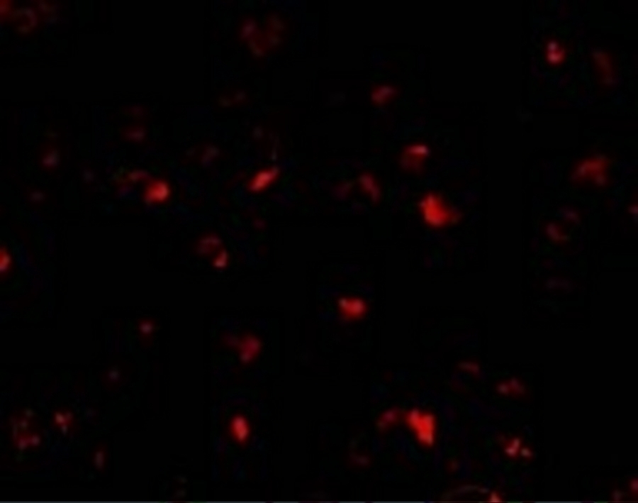
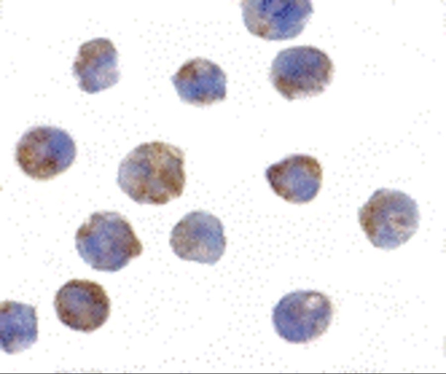
Antibody validated: Western Blot in human, mouse and rat samples; Immunocytochemistry in human samples; Immunofluorescence in human samples. All other applications and species not yet tested.
Specificity:
This polyclonal antibody has no cross-reaction to PHAP I2a and PHAP III.
Positive Control 1:
Cat. No. 1207 - Raji Cell Lysate
Positive Control 2:
Cat. No. 17-007 - Raji Cell Slide
Predicted Molecular Weight:
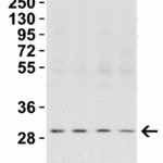
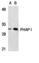
Predicted: 29kD
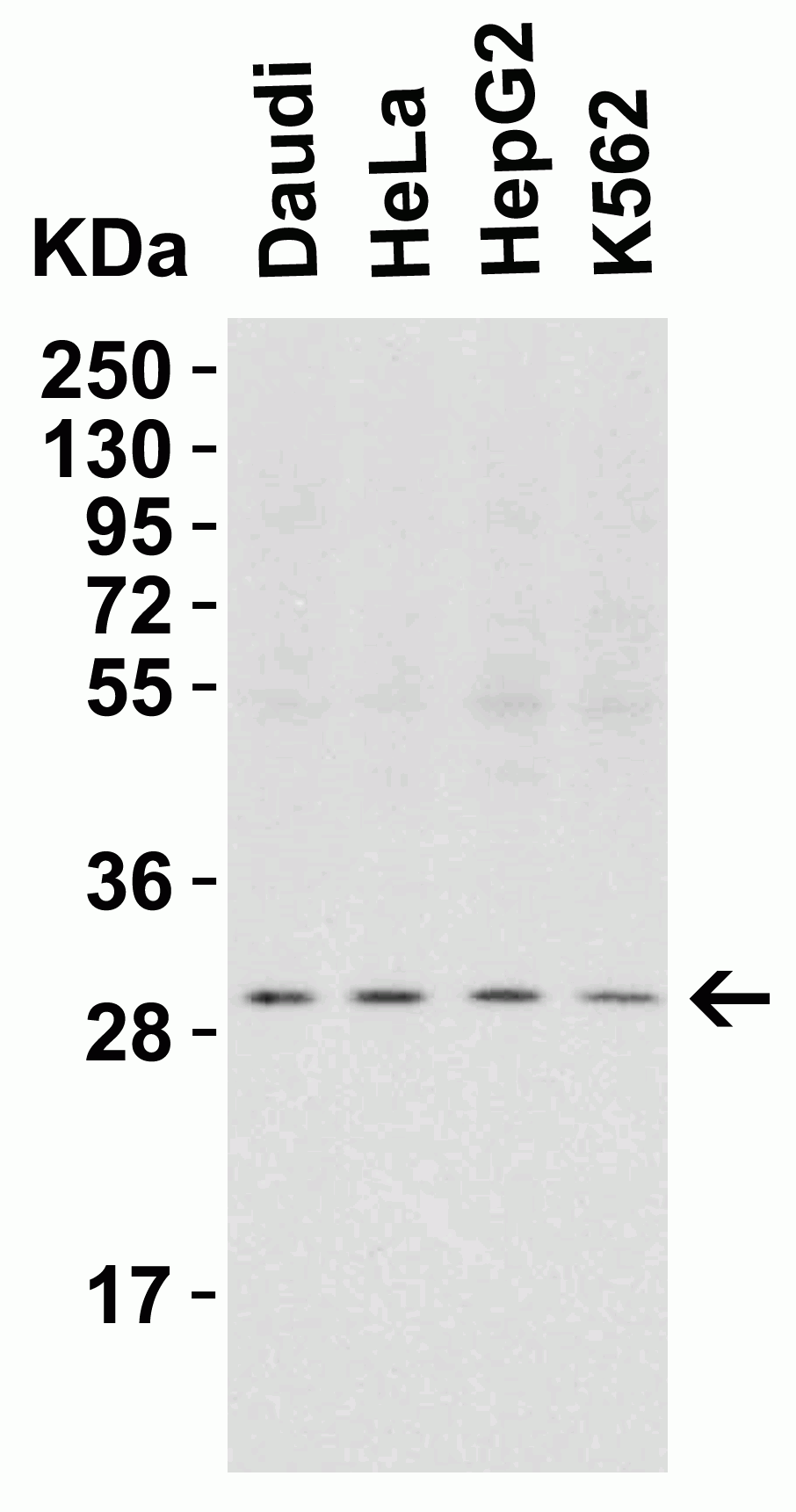
Observed: 29 kD
Validation:

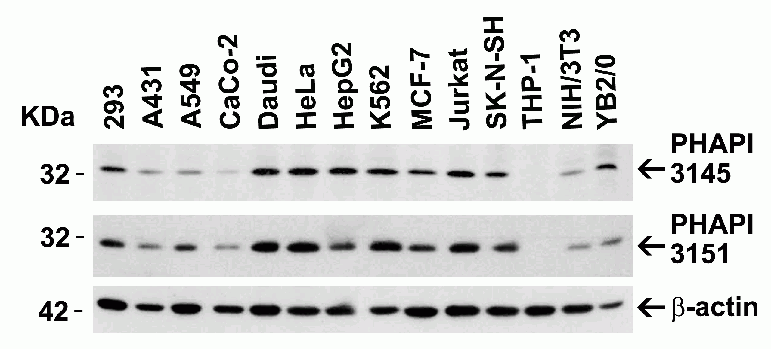
Independent Antibody Validation in Cell lines (Figure 2) shows similar PHAP I expression profile in human, mouse and rat cell lines detected by two independent anti-PHAP I antibodies that recognize different epitopes, 3145 against C-terminus domain and 3151 against C-terminus different domain. PHAP I proteins are detected in the most tested cell lines at different expression levels by the two independent antibodies.

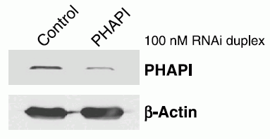
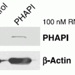
KD validation (Figure 6): Anti-PHAP I antibody (3151) specificity was further verified by PHAP I specific knockdown. PHAP I signal in T47D cells transfected with PHAP I siRNAs was disrupted in comparison with that in cells transfected with control siRNAs.

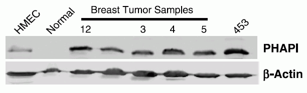
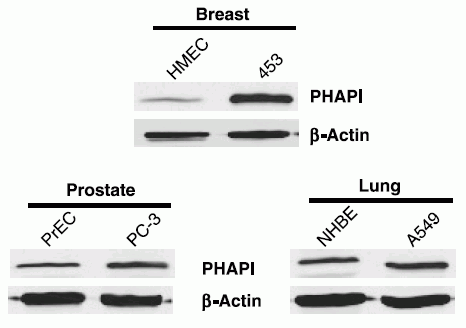
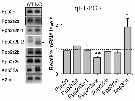
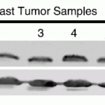
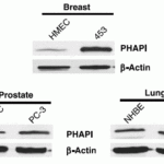
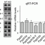
Regulated expression validation (Figure 7, 8, 9): PHAP I expression detected by anit-PHAP I antibodies (3151) was up-regulated in breast cancer tissues of patients (Figure 7) and the breast cancer cell line (Figure 8) as well as in the Atxn1 KO mice (Figure 9).

Isoforms:
Human PHAP I has one isoform (249aa, 28.6kD). Mouse PHAP I has one isoform (247aa, 28.5kD) and rat PHAP I also has one isoform (247aa, 28.6kD). 3151 can detect all isoforms of human, mouse and rat.
Purification:
PHAP I Antibody is DEAE purified.
Clonality:
Polyclonal
Isotype:
IgG
Physical State:
Liquid
Buffer:
PHAP I Antibody is supplied in PBS containing ≤0.09% sodium azide.
Concentration:
1 mg/mL
Storage Conditions:
PHAP I antibody can be stored at 4°C, stable for up to one year. Antibodies should not be exposed to prolonged high temperatures.
Ncbi Official Symbol:
ANP32A
Additional Names:
PHAP I Antibody: LANP, MAPM, PP32, HPPCn, PHAP1, PHAPI, I1PP2A, C15orf1, LANP, Acidic leucine-rich nuclear phosphoprotein 32 family member A, Acidic nuclear phosphoprotein pp32
Protein Accession Number:
P39687
Protein Gi Number:
730318
Ncbi Gene Id Number:
8125
User Note:
Optimal dilutions for each application to be determined by the researcher.
Background:
PHAP I Antibody: Apoptosis is related to many diseases and development. Caspase-9 plays a central role in cell death induced by a variety of apoptosis activators. Cytochrome c, after released from mitochondria, binds to Apaf-1, which forms an apoptosome that in turn binds to and activate procaspase-9. Activated caspase-9 cleaves and activates the effector caspases (caspase-3, -6 and -7), which are responsible for the proteolytic cleavage of many key proteins in apoptosis. The tumor suppressor putative HLA-DR-associated proteins (PHAPs) were recently identified as important regulators of mitochondrion apoptosis. PHAP appears to facilitate apoptosome-medicated caspase-9 activation and to stimulate the mitochondrial apoptotic pathway. PHAP was also shown to oppose both Ras- and Myc-medicated cell transformation.
Background Reference 1:
Jiang et al. Science. 2003;299(5604):223-6.
Background Reference 2:
Nicholson and Thornberry. Science. 2003 10;299(5604):214-5.
Citation 1:
Schafer et al. Enhanced sensitivity to cytochrome c-induced apoptosis mediated by PHAPI in breast cancer cells. Cancer Res. 2006;66(4):2210-8. PMID: 16489023
Citation 2:
Sánchez et al. A novel function of Ataxin-1 in the modulation of PP2A activity is dysregulated in the spinocerebellar ataxia type 1. Hum Mol Genet. 2013 22(17):3425-37. PMID: 23630944
Citation 3:
Kurokawa et al. A network of substrates of the E3 ubiquitin ligases MDM2 and HUWE1 control apoptosis independently of p53. Sci Signal. 2013;6(274):ra32. PMID: 23652204
Citation 4:
Parrish. Regulation of Apoptosis Following Mitochondrial Cytochrome c Release. Department of Pharmacology and Molecular Cancer Biology, Duke University. PhD Thesis, 2010.

FOR RESEARCH USE ONLY

For additional information, visit ProSci’s Terms & Conditions Page.

Disclaimer:
Optimal dilutions/concentrations should be determined by the end user. The information provided is a guideline for product use. This product is for research use only.

CATALOG NUMBER:

3145

List Size:
0.02 mg, 0.1 mg

List Price:

Price range: $99.00 through $445.00

Shipping Info
Customize your order
Custom Antibody Services

New & Featured Products

CATALOG NUMBER: PM-7369-HRP

CATALOG NUMBER: PM-7369-biotin

CATALOG NUMBER: PM-7369