Most Cited
Trial Size

SARS-CoV-2 (COVID-19) Envelope Antibody

CATALOG NUMBER: 3531

Clonality:
Polyclonal
Tested Applications:
ELISA, IF, IHC
Host Species:
Rabbit
Species Reactivity:
Virus
Conjugate:
Unconjugated
Specifications
Host Species:
Rabbit
Species Reactivity:
Virus
Homology:
Predicted reactivity based on immunogen sequence: SARS-CoV Envelope proteins: (100%)
Immunogen:
Anti-SARS-CoV-2 (COVID-19, 2019-nCoV) Envelope antibody (3531) was raised against a peptide corresponding to 10 amino acids near the amino terminus of SARS-CoV-2 (COVID-19, 2019-nCoV) Envelope protein.
The immunogen is located within the first 50 amino acids of SARS-CoV-2 (COVID-19, 2019-nCoV) Envelope.
Conjugate:
Unconjugated
Tested Applications:
ELISA, IF, IHC
Application Note:
IHC/IF: 1-3 μg/mL
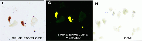
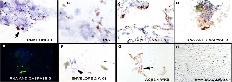
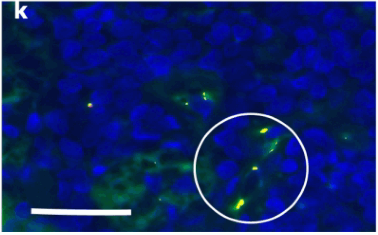
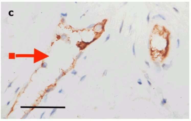
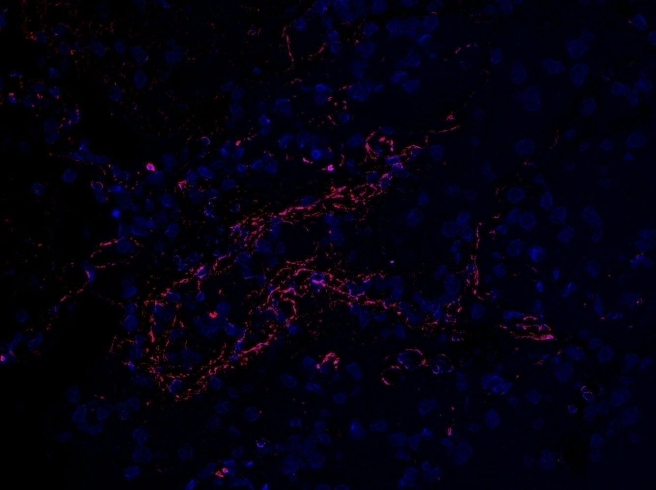
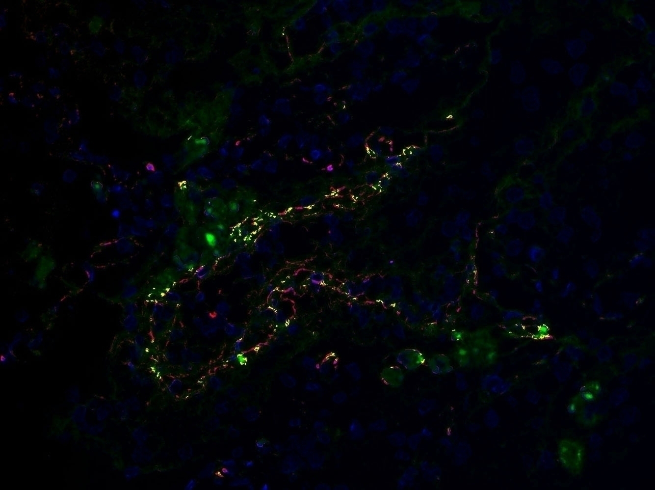
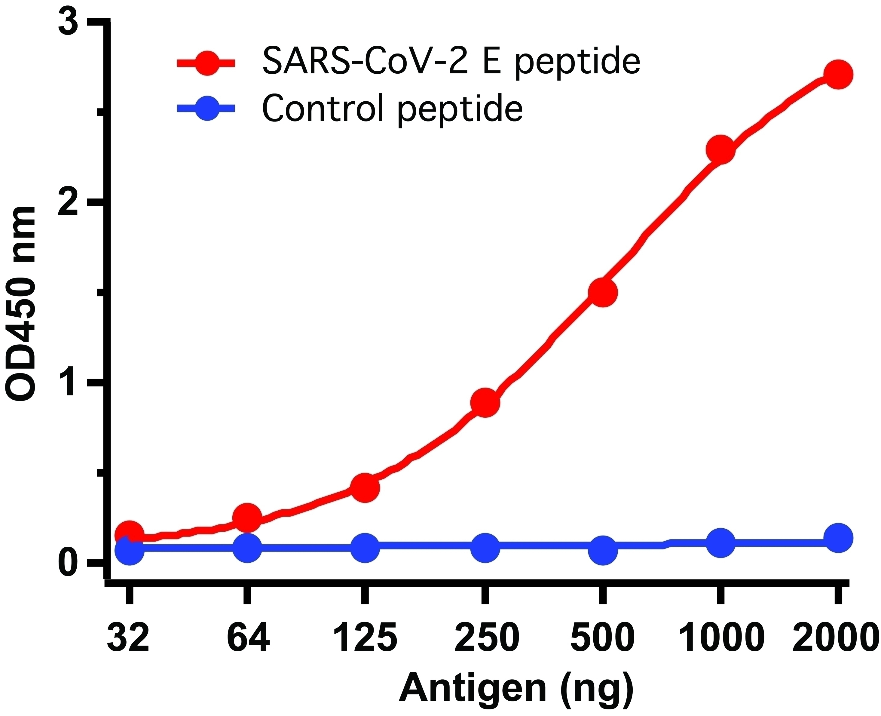
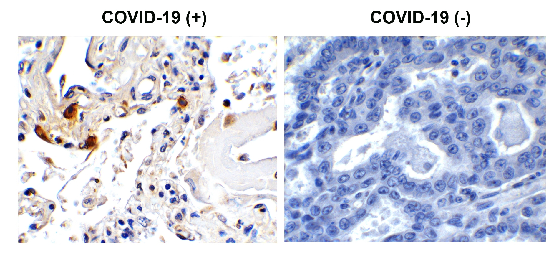
Antibody validated: Immunohisochemistry and immunofuorescence in COVID-19 patient samples. SARS-CoV-2 (COVID-19, 2019-nCoV) Envelope antibody can detect 50 ng of free peptide at 1 μg/mL in ELISA. But it cannot detect envelope recombinant protein in WB and ELISA.
Isoforms:
SARS-CoV-2 (COVID-19, 2019-nCoV) Envelope has one isoform (75aa).
Purification:
SARS-CoV-2 (COVID-19, 2019-nCoV) Envelope Antibody is affinity chromatography purified via peptide column.
Clonality:
Polyclonal
Isotype:
IgG
physical-state:
Liquid
Buffer:
SARS-CoV-2 (COVID-19, 2019-nCoV) Envelope Antibody is supplied in PBS containing 0.02% sodium azide.
Concentration:
1 mg/mL
Storage Conditions:
SARS-CoV-2 (COVID-19, 2019-nCoV) Envelope antibody can be stored at 4 ° C for three months and -20 ° C, stable for up to one year. As with all antibodies care should be taken to avoid repeated freeze thaw cycles. Antibodies should not be exposed to prolonged high temperatures.
Ncbi Official Symbol:
E
Additional Names:
SARS-CoV-2 (COVID-19, 2019-nCoV) Envelope Antibody: Severe acute respiratory syndrome coronavirus 2 (SARS-CoV-2), Envelope protein, E protein
Protein Accession Number:
QHD43418
Protein Gi Number:
1791269092
Ncbi Gene Id Number:
43740570
User Note:
Optimal dilutions for each application to be determined by the researcher.
Background:
Coronavirus disease 2019 (COVID-19), formerly known as 2019-nCoV acute respiratory disease, is an infectious disease caused by SARS-CoV-2, a virus closely related to the SARS virus (1). The disease is the cause of the 2019–20 coronavirus outbreak (2). The structure of 2019-nCoV consists of the following: a spike protein (S), hemagglutinin-esterease dimer (HE), a membrane glycoprotein (M), an envelope protein (E) a nucleoclapid protein (N) and RNA. Envelope protein is a small polypeptide that contains at least one alpha-helical transmembrane domain. It involves in several aspects of the virus's life cycle, such as assembly, budding, envelope formation, and pathogenesis. E protein has membrane permeabilizing activity, which provides a possible rationale to inhibit in vitro ion channel activity of some synthetic coronavirus E proteins, and also viral replication (3).

Background Reference 1:
Gorbalenya. bioRxiv: 2020.
Background Reference 2:
Hui et al. Int J Infect Dis. 2020;91:264-266.
Background Reference 3:
Pervushin et al. PLoS Pathog. 2009; 5(7): e1000511.
Citation 1:
Nuovo GJ et al. Cytologic and molecular correlates of SARS-CoV-2 infection of the nasopharynx. Ann Diagn Pathol. 2020; 48:151565. doi: 10.1016/j.anndiagpath.2020.151565. PMID: 32659620
Citation 2:
Magro C, Mulvey JJ, Laurence J, Sanders S, Crowson N, Grossman M, Harp J, Nuovo G. The differing pathophysiologies that underlie COVID-19 associated perniosis and thrombotic retiform purpura: a case series. Br J Dermatol. 2020; 22:10.1111/bjd.19415. doi: 10.1111/bjd.19415. PMID: 32779733
Citation 3:
Nuovo G, Tili E, Suster D, Matys E, Hupp L, Magro C. Strong homology between SARS-CoV-2 envelope protein and a Mycobacterium sp. antigen allows rapid diagnosis of Mycobacterial infections and may provide specific anti-SARS-CoV-2 immunity via the BCG vaccine. Ann Diagn Pathol. 2020; 48:151600. doi: 10.1016/j.anndiagpath.2020.151600. PMID: 32805515
Citation 4:
Ko, et al. Discordant anti-SARS-CoV-2 spike protein and RNA staining in cutaneous perniotic lesions suggests endothelial deposition of cleaved spike protein. J Cutan Pathol . 2021 Jan;48(1):47-52. doi: 10.1111/cup.13866. Epub 2020 Oct 1.PMID: 32895985 
Citation 5:
Magro, et al. Docked severe acute respiratory syndrome coronavirus 2 proteins within the cutaneous and subcutaneous microvasculature and their role in the pathogenesis of severe coronavirus disease 2019. Hum Pathol. 2020 Dec;106:106-116. doi: 10.1016/j.humpath.2020.10.002. Epub 2020 Oct 12.PMID: 33058948
Citation 6:
Magro, et al. The differing pathophysiologies that underlie COVID-19-associated perniosis and thrombotic retiform purpura: a case series. Br J Dermatol. 2020 Jul 22;10.1111/bjd.19415. doi: 10.1111/bjd.19415PMID: 32779733
Citation 7:
Mulvey, et al. Analysis of complement deposition and viral RNA in placentas of COVID-19 patients. Ann Diagn Pathol. 2020 Jun;46:151530. doi: 10.1016/j.anndiagpath.2020.151530. Epub 2020 Apr 25.PMID: 32387855
Citation 8:
Rotman, et al. Concomitant calciphylaxis and COVID-19 associated thrombotic retiform purpura. Skeletal Radiol. 2020 Nov;49(11):1879-1884. doi: 10.1007/s00256-020-03579-6. Epub 2020 Aug 26.PMID: 32844243

FOR RESEARCH USE ONLY

For additional information, visit ProSci’s Terms & Conditions Page.

Disclaimer:
Optimal dilutions/concentrations should be determined by the end user. The information provided is a guideline for product use. This product is for research use only.

CATALOG NUMBER:

3531

List Size:
0.02 mg, 0.1 mg

List Price:

Price range: $99.00 through $445.00

Shipping Info
Customize your order
Custom Antibody Services

New & Featured Products

CATALOG NUMBER: PM-7369-HRP

CATALOG NUMBER: PM-7369-biotin

CATALOG NUMBER: PM-7369