Search Our Antibody Catalog

Browse thousands of high-quality, US-produced antibodies and reagents for research, diagnostics, and development.

Host Species
Applications
Conjugate
Clone Type
Product Type
Research Area
Species Reactivity
Modifications
Product Badges
Product Badges
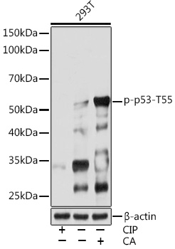
Western blot analysis of lysates from 293T cells, using Phospho-p53-T55 Rabbit mAb at 1:1000 dilution. 293T cells were treated with CIP(20uL/400ul) at 37℃ for 1 hour or treated with Calyculin A (100 nM) at 37℃ for 30 minutes after serum-starvation overnight.|Secondary antibody: HRP-conjugated Goat anti-Rabbit IgG (H+L) at 1:10000 dilution.|Lysates/proteins: 25μg per lane.|Blocking buffer: 3% BSA.|Detection: ECL Basic Kit.|Exposure time: 30s.
Clonality:
Monoclonal
Host Species:
Rabbit
Tested Applications:
ELISA, WB
Conjugate:
Unconjugated
Species Reactivity:
Human
List Size:
100 uL
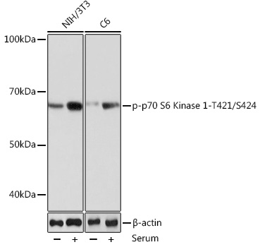
Western blot analysis of various lysates using Phospho-p70 S6 Kinase 1-T421/S424 Rabbit mAb at 1:1000 dilution. NIH/3T3 and C6 cells were treated with 10% FBS at 37℃ for 30 minutes after serum-starvation overnight|Secondary antibody: HRP-conjugated Goat anti-Rabbit IgG (H+L) at 1:10000 dilution.|Lysates/proteins: 25μg per lane.|Blocking buffer: 3% nonfat dry milk in TBST.|Detection: ECL Basic Kit.|Exposure time: 60S.
Clonality:
Monoclonal
Host Species:
Rabbit
Tested Applications:
ELISA, IF/ICC, WB
Conjugate:
Unconjugated
Species Reactivity:
Human, Mouse, Rat
List Size:
100 uL
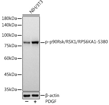
Western blot analysis of lysates from NIH/3T3 cells, using Phospho-p90Rsk/RSK1/RPS6KA1-S380 Rabbit mAb at 1:1000 dilution. NIH/3T3 cells were treated with PDGF (100 ng/ml) at 37℃ for 30 minutes after serum-starvation overnight.|Secondary antibody: HRP-conjugated Goat anti-Rabbit IgG (H+L) at 1:10000 dilution.|Lysates/proteins: 25μg per lane.|Blocking buffer: 3% nonfat dry milk in TBST.|Detection: ECL Basic Kit.|Exposure time: 3s.
Clonality:
Monoclonal
Host Species:
Rabbit
Tested Applications:
ELISA, WB
Conjugate:
Unconjugated
Species Reactivity:
Human, Mouse, Rat
List Size:
100 uL
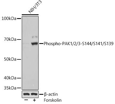
Western blot analysis of various lysates using Phospho-PAK1/2/3-S144/S141/S139 Rabbit mAb at 1:1000 dilution. NIH/3T3 cells were treated with Forskolin (30 uM) at 37℃ for 30 minutes after serum-starvation overnight|Secondary antibody: HRP-conjugated Goat anti-Rabbit IgG (H+L) at 1:10000 dilution.|Lysates/proteins: 25μg per lane.|Blocking buffer: 3% BSA.|Detection: ECL Basic Kit.|Exposure time: 1s.
Clonality:
Monoclonal
Host Species:
Rabbit
Tested Applications:
ELISA, WB
Conjugate:
Unconjugated
Species Reactivity:
Human, Mouse, Rat
List Size:
100 uL
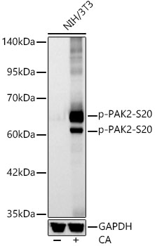
Western blot analysis of lysates from NIH/3T3 cells, using Phospho-PAK2-S20 Rabbit mAb at 1:2000 dilution. NIH/3T3 cells were treated with Calyculin A (100 nM) at 37℃ for 30 minutes after serum-starvation overnight.|Secondary antibody: HRP-conjugated Goat anti-Rabbit IgG (H+L) at 1:10000 dilution.|Lysates/proteins: 25μg per lane.|Blocking buffer: 3% nonfat dry milk in TBST.|Detection: ECL Basic Kit.|Exposure time: 10s.
Clonality:
Monoclonal
Host Species:
Rabbit
Tested Applications:
ELISA, WB
Conjugate:
Unconjugated
Species Reactivity:
Mouse, Rat
List Size:
100 uL
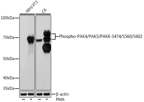
Western blot analysis of various lysates using Phospho-PAK4/PAK5/PAK6-S474/S560/S602 Rabbit mAb at 1:1000 dilution. Both NIH/3T3 cells and C6 cells were treated with PMA/TPA (200 nM) at 37℃ for 30 minutes after serum-starvation overnight.|Secondary antibody: HRP-conjugated Goat anti-Rabbit IgG (H+L) at 1:10000 dilution.|Lysates/proteins: 25μg per lane.|Blocking buffer: 3% BSA.|Detection: ECL Basic Kit.|Exposure time: 10s.
Clonality:
Monoclonal
Host Species:
Rabbit
Tested Applications:
ELISA, WB
Conjugate:
Unconjugated
Species Reactivity:
Mouse, Rat
List Size:
100 uL
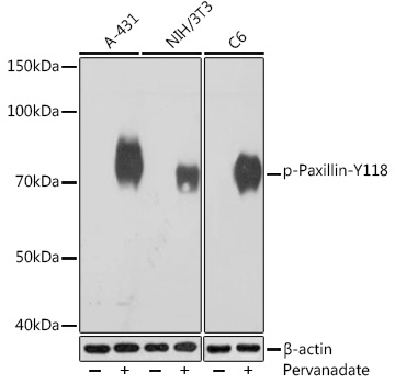
Western blot analysis of various lysates using Phospho-Paxillin-Y118 Rabbit mAb at 1:1000 dilution. A-431 cells were treated with pervanadate (1 mM) at 37℃ for 30 minutes after serum-starvation overnight.  NIH/3T3 cells were treated with pervanadate (1 mM) at 37℃ for 30 minutes after serum-starvation overnight. C6 cells were treated with pervanadate (1 mM) at 37℃ for 30 minutes after serum-starvation overnight.|Secondary antibody: HRP-conjugated Goat anti-Rabbit IgG (H+L) at 1:10000 dilution.|Lysates/proteins: 25μg per lane.|Blocking buffer: 3% BSA.|Detection: ECL Basic Kit.|Exposure time: 1s.
Clonality:
Monoclonal
Host Species:
Rabbit
Tested Applications:
ELISA, WB
Conjugate:
Unconjugated
Species Reactivity:
Human, Mouse, Rat
List Size:
100 uL
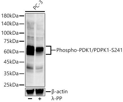
Western blot analysis of various lysates, using Phospho-PDK1/PDPK1-S241 Rabbit mAb at 1:2000 dilution. PC-3 cells were treated by λ-PP mixed solution (1ul) at 30℃ for 30 minutes.|Secondary antibody: HRP-conjugated Goat anti-Rabbit IgG (H+L) at 1:10000 dilution.|Lysates/proteins: 25μg per lane.|Blocking buffer: 3% nonfat dry milk in TBST.|Detection: ECL Enhanced Kit.|Exposure time: 120s.
Clonality:
Monoclonal
Host Species:
Rabbit
Tested Applications:
ELISA, WB
Conjugate:
Unconjugated
Species Reactivity:
Human
List Size:
100 uL
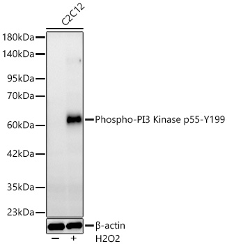
Western blot analysis of lysates from C2C12 cells using Phospho-PI3 Kinase p55-Y199 Rabbit mAb at 1:5000 dilution. C2C12 cells were treated with Hydrogen Peroxide (2 nM) at 37℃ for 15 minutes after serum-starvation overnight.|Secondary antibody: HRP-conjugated Goat anti-Rabbit IgG (H+L) at 1:10000 dilution.|Lysates/proteins: 25 μg per lane.|Blocking buffer: 3% nonfat dry milk in TBST.|Detection: ECL Basic Kit.|Exposure time: 60s.
Clonality:
Monoclonal
Host Species:
Rabbit
Tested Applications:
ELISA, IF/ICC, WB
Conjugate:
Unconjugated
Species Reactivity:
Mouse
List Size:
100 uL
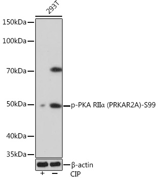
Western blot analysis of lysates from 293T cells, using Phospho-PKA RIIα (PRKAR2A)-S99 Rabbit mAb at 1:1000 dilution. 293T cells were treated by CIP(20uL/400ul) at 37℃ for 1 hour.|Secondary antibody: HRP-conjugated Goat anti-Rabbit IgG (H+L) at 1:10000 dilution.|Lysates/proteins: 25μg per lane.|Blocking buffer: 3% BSA.|Detection: ECL Basic Kit.|Exposure time: 90s.
Clonality:
Monoclonal
Host Species:
Rabbit
Tested Applications:
ELISA, WB
Conjugate:
Unconjugated
Species Reactivity:
Human, Rat
List Size:
100 uL