Amyloid Plaque and Neurofibrillary Tangle Formation in Alzheimer's Disease

Product Type
Research Area
Clonality
Host Species
Species Reactivity
Tested Applications
Conjugate
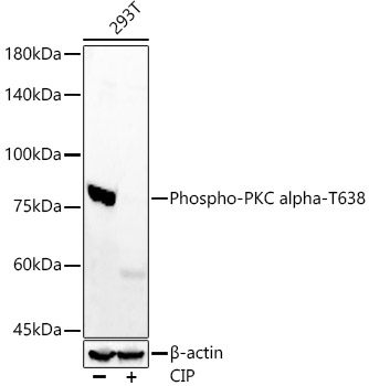
Western blot analysis of lysates from 293T cells, using Phospho-PKC alpha-T638 Rabbit mAb at  1:7000 dilution. 293T cells were treated by CIP(20uL/400ul) at 37℃ for 1 hour.|Secondary antibody: HRP-conjugated Goat anti-Rabbit IgG (H+L) at 1:10000 dilution.|Lysates/proteins: 25μg per lane.|Blocking buffer: 3% nonfat dry milk in TBST.|Detection: ECL Enhanced Kit.|Exposure time: 180s.
Clonality:
Monoclonal
Host Species:
Rabbit
Tested Applications:
ELISA, WB
Conjugate:
Unconjugated
Species Reactivity:
Human
List Size:
100 uL
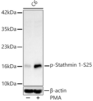
Western blot analysis of lysates from C6 cells, using Phospho-Stathmin 1-S25 Rabbit mAb at 1:1000 dilution. C6 cells were treated with PMA/TPA (200 nM) at 37℃ for 30 minutes after serum-starvation overnight.|Secondary antibody: HRP-conjugated Goat anti-Rabbit IgG (H+L) at 1:10000 dilution.|Lysates/proteins: 25μg per lane.|Blocking buffer: 3% nonfat dry milk in TBST.|Detection: ECL Basic Kit.|Exposure time: 90s.
Clonality:
Monoclonal
Host Species:
Rabbit
Tested Applications:
ELISA, WB
Conjugate:
Unconjugated
Species Reactivity:
Human, Rat
List Size:
100 uL
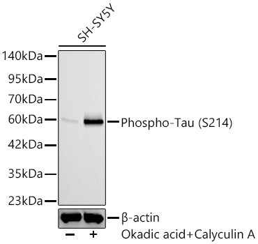
Western blot analysis lysates from SH-SY5Y, using Phospho-Tau (S214) Rabbit mAb at 1:1000 dilution. SH-SY5Y cells were treated with Okadaic Acid (100 nM) at 37℃ for 1 hour and Calyculin A (100nM) at 37℃ for 30 minutes after serum-starvation overnight.℃ for 30 minutes after serum-starvation overnight.|Secondary antibody: HRP-conjugated Goat anti-Rabbit IgG (H+L) at 1:10000 dilution.|Lysates/proteins: 25ug per lane.|Blocking buffer: 3% nonfat dry milk in TBST.|Detection: ECL Basic Kit.|Exposure time: 60s.
Clonality:
Monoclonal
Host Species:
Rabbit
Tested Applications:
ELISA, IHC-P, WB
Conjugate:
Unconjugated
Species Reactivity:
Human, Mouse
List Size:
100 uL
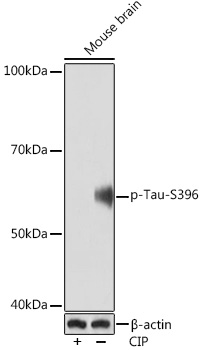
Western blot analysis of lysates from Mouse brain, using Phospho-Tau-S396 Rabbit mAb at 1:1000 dilution. Mouse brain were treated by CIP(20μL/400μL) at 37℃ for 1 hour.|Secondary antibody: HRP-conjugated Goat anti-Rabbit IgG (H+L) at 1:10000 dilution.|Lysates/proteins: 25μg  per lane.|Blocking buffer: 3% BSA.|Detection: ECL Basic Kit.|Exposure time: 90s.
Clonality:
Monoclonal
Host Species:
Rabbit
Tested Applications:
ELISA, WB
Conjugate:
Unconjugated
Species Reactivity:
Mouse
List Size:
100 uL
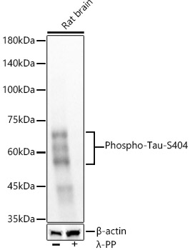
Western blot analysis of lysates from rat brain, using Phospho-Tau-S404 Rabbit mAb at  1:5000 dilution. Rat brain cells were treated with λ-PP mixed solution (1ul) at 30℃ for 30 minutes.|Secondary antibody: HRP-conjugated Goat anti-Rabbit IgG (H+L) at 1:10000 dilution.|Lysates/proteins: 25μg per lane.|Blocking buffer: 3% nonfat dry milk in TBST.|Detection: ECL Basic Kit.|Exposure time: 10s.
Clonality:
Monoclonal
Host Species:
Rabbit
Tested Applications:
ELISA, WB
Conjugate:
Unconjugated
Species Reactivity:
Human, Mouse, Rat
List Size:
100 uL
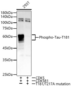
Western blot analysis of lysates from 293T cells transfected with CDK5+CDK5R1+Tau and  CDK5+CDK5R1+mutant Tau(T181/T217A) using Phospho-Tau-T181 Rabbit mAb at1:20000 dilution.|Secondary antibody: HRP-conjugated Goat anti-Rabbit IgG (H+L) at 1:10000 dilution.|Lysates/proteins: 25μg per lane.|Blocking buffer: 3% nonfat dry milk in TBST.|Detection: ECL Basic Kit.|Exposure time: 10s.
Clonality:
Monoclonal
Host Species:
Rabbit
Tested Applications:
ELISA, WB
Conjugate:
Unconjugated
Species Reactivity:
Human
List Size:
100 uL
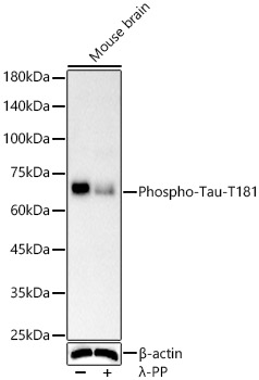
Western blot analysis of various lysates, using Phospho-Tau-T181 antibody at  1:2000 dilution. Mouse brain were treated by λ-PP mixed solution (1ul) at 30℃ for 30 minutes.|Secondary antibody: HRP-conjugated Goat anti-Rabbit IgG (H+L) at 1:10000 dilution.|Lysates/proteins: 25μg  per lane.|Blocking buffer: 3% nonfat dry milk in TBST.|Detection: ECL Enhanced Kit.|Exposure time: 60s.
Clonality:
Monoclonal
Host Species:
Rabbit
Tested Applications:
ELISA, IF-P, IHC-P, IP, WB
Conjugate:
Unconjugated
Species Reactivity:
Human, Mouse, Rat
List Size:
100 uL
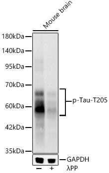
Western blot analysis of lysates from Mouse brain, using Phospho-Tau-T205 Rabbit mAb at 1:1000 dilution.|Secondary antibody: HRP-conjugated Goat anti-Rabbit IgG (H+L) at 1:10000 dilution.|Lysates/proteins: 25μg per lane.|Blocking buffer: 3% nonfat dry milk in TBST.|Detection: ECL Basic Kit.|Exposure time: 20s.
Clonality:
Monoclonal
Host Species:
Rabbit
Tested Applications:
ELISA, IF-P, IHC-P, WB
Conjugate:
Unconjugated
Species Reactivity:
Mouse, Rat
List Size:
100 uL
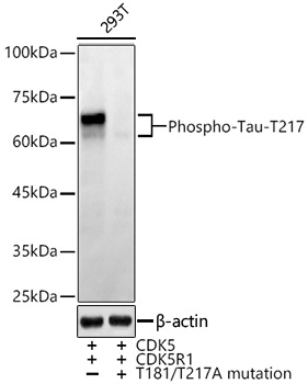
Western blot analysis of lysates from from 293T cells transfected with CDK5+CDK5R1+Tau and CDK5+CDK5R1+mutant Tau(T181/T217A) using Phospho-Tau-T217 Rabbit mAb at  1:10000 dilution.|Secondary antibody: HRP-conjugated Goat anti-Rabbit IgG (H+L) at 1:10000 dilution.|Lysates/proteins: 25μg per lane.|Blocking buffer: 3% nonfat dry milk in TBST.|Detection: ECL Enhanced Kit.|Exposure time: 60s.
Clonality:
Monoclonal
Host Species:
Rabbit
Tested Applications:
ELISA, WB
Conjugate:
Unconjugated
Species Reactivity:
Human
List Size:
100 uL
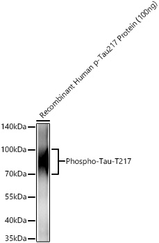
Western blot analysis of recombinant Human p-Tau217 Protein using Phospho-Tau-T217 Rabbit mAb at 1:1000 dilution incubated overnight at 4℃.|Secondary antibody: HRP-conjugated Goat anti-Rabbit IgG (H+L) at 1:10000 dilution.|Lysates/proteins: 100ng μg per lane.|Blocking buffer: 3% nonfat dry milk in TBST.|Detection: ECL Basic Kit.|Exposure time: 5s.
Clonality:
Monoclonal
Host Species:
Rabbit
Tested Applications:
ELISA, IF/ICC, WB
Conjugate:
Unconjugated
Species Reactivity:
Human, Mouse
List Size:
100 uL