We use cookies on our website to give you the most relevant experience by remembering your preferences and repeat visits. By clicking “Accept”, you consent to the use of cookies.
This website uses cookies to improve your experience while you navigate through the website. Out of these, the cookies that are categorized as necessary are stored on your browser as they are essential for the working of basic functionalities of the website. We also use third-party cookies that help us analyze and understand how you use this website. These cookies will be stored in your browser only with your consent. You also have the option to opt-out of these cookies. But opting out of some of these cookies may affect your browsing experience.
Necessary cookies are absolutely essential for the website to function properly. These cookies ensure basic functionalities and security features of the website, anonymously.
Cookie
Duration
Description
cookielawinfo-checbox-analytics
11 months
This cookie is set by GDPR Cookie Consent plugin. The cookie is used to store the user consent for the cookies in the category "Analytics".
cookielawinfo-checbox-functional
11 months
The cookie is set by GDPR cookie consent to record the user consent for the cookies in the category "Functional".
cookielawinfo-checbox-others
11 months
This cookie is set by GDPR Cookie Consent plugin. The cookie is used to store the user consent for the cookies in the category "Other.
cookielawinfo-checkbox-necessary
11 months
This cookie is set by GDPR Cookie Consent plugin. The cookies is used to store the user consent for the cookies in the category "Necessary".
cookielawinfo-checkbox-performance
11 months
This cookie is set by GDPR Cookie Consent plugin. The cookie is used to store the user consent for the cookies in the category "Performance".
viewed_cookie_policy
11 months
The cookie is set by the GDPR Cookie Consent plugin and is used to store whether or not user has consented to the use of cookies. It does not store any personal data.
Functional cookies help to perform certain functionalities like sharing the content of the website on social media platforms, collect feedbacks, and other third-party features.
Performance cookies are used to understand and analyze the key performance indexes of the website which helps in delivering a better user experience for the visitors.
Analytical cookies are used to understand how visitors interact with the website. These cookies help provide information on metrics the number of visitors, bounce rate, traffic source, etc.
Advertisement cookies are used to provide visitors with relevant ads and marketing campaigns. These cookies track visitors across websites and collect information to provide customized ads.

![Western blot analysis of lysates from wild type (WT) and BRD2 knockdown (KD) 293T cells using [KD Validated] BRD2 Rabbit mAb at 1:1000 dilution incubated at room temperature for 1.5 hours.|Secondary antibody: HRP-conjugated Goat anti-Rabbit IgG (H+L) at 1:10000 dilution.|Lysates/proteins: 25 μg per lane.|Blocking buffer: 3% nonfat dry milk in TBST.|Detection: ECL Basic Kit.|Exposure time: 90s.](https://www.prosciantibodies.com/wp-content/uploads/products/ARC56992-01_WB_02_82-493.jpg)
![Western blot analysis of lysates from Hep G2 cells using [KD Validated] ROMO1 Rabbit mAb at 1:14000 dilution incubated at room temperature for 1.5 hours.|Secondary antibody: HRP-conjugated Goat anti-Rabbit IgG (H+L) at 1:10000 dilution.|Lysates/proteins: 25 μg per lane.|Blocking buffer: 3% nonfat dry milk in TBST.|Detection: ECL Basic Kit.|Exposure time: 10s.](https://www.prosciantibodies.com/wp-content/uploads/products/ARC71752-01_WB_01_83-528.jpg)
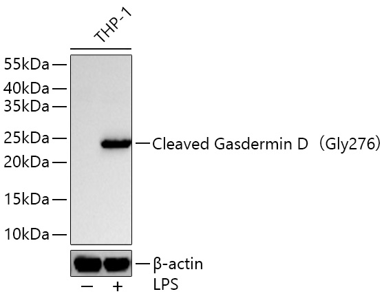
![Western blot analysis of lysates from THP-1 cells using [KO Validated] Cleaved Gasdermin D (N Terminal) Rabbit mAb at 1:72000 dilution incubated overnight at 4℃. THP-1 cells were treated with PMA/TPA (80 nM) at 37℃ for overnight and LPS (1 μg/ml) at 37℃ for 6 hours. | Secondary antibody: HRP-conjugated Goat anti-Rabbit IgG (H+L) at 1:10000 dilution.|Lysates/proteins: 30 μg per lane.| Blocking buffer: 3 % nonfat dry milk in TBST.| Detection: ECL Basic Kit.| Exposure time: 45s.](https://www.prosciantibodies.com/wp-content/uploads/products/ARC57989-03_WB_01_82-855.jpg)