
Hedgehog Signaling Pathway

Product Type
Clonality
Host Species
Species Reactivity
Tested Applications
Conjugate
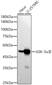
Western blot analysis of various lysates using GSK-3α/β Rabbit mAb at1:2000 dilution.|Secondary antibody: HRP-conjugated Goat anti-Rabbit IgG (H+L) at 1:10000 dilution.|Lysates/proteins: 25μg per lane.|Blocking buffer: 3% nonfat dry milk in TBST.|Detection: ECL Basic Kit.|Exposure time: 180s.
Clonality:
Monoclonal
Host Species:
Rabbit
Tested Applications:
ELISA, WB
Conjugate:
Unconjugated
Species Reactivity:
Human, Rat
List Size:
100 uL
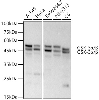
Western blot analysis of various lysates, using GSK-3α/β Rabbit mAb at 1:20000 dilution.|Secondary antibody: HRP-conjugated Goat anti-Rabbit IgG (H+L) at 1:10000 dilution.|Lysates/proteins: 25μg per lane.|Blocking buffer: 3% nonfat dry milk in TBST.|Detection: ECL Basic Kit.|Exposure time: 90s.
Clonality:
Monoclonal
Host Species:
Rabbit
Tested Applications:
ELISA, IP, WB
Conjugate:
Unconjugated
Species Reactivity:
Human, Mouse, Rat
List Size:
100 uL
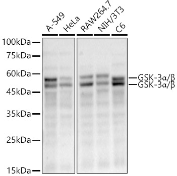
Western blot analysis of various lysates, using GSK-3α/β Rabbit mAb at 1:50000 dilution.|Secondary antibody: HRP-conjugated Goat anti-Rabbit IgG (H+L) at 1:10000 dilution.|Lysates/proteins: 25μg per lane.|Blocking buffer: 3% nonfat dry milk in TBST.|Detection: ECL Enhanced Kit.|Exposure time: 90s.
Clonality:
Monoclonal
Host Species:
Rabbit
Tested Applications:
ELISA, IF-P, IF/ICC, IHC-P, WB
Conjugate:
Unconjugated
Species Reactivity:
Human, Mouse, Rat
List Size:
100 uL
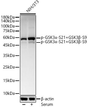
Western blot analysis of lysates from NIH/3T3 cells, using Phospho-GSK3α-S21+GSK3β-S9 Rabbit mAb at  1:800 dilution. NIH/3T3 cells were treated with 10% FBS at 37℃ for 30 minutes after serum-starvation overnight.|Secondary antibody: HRP-conjugated Goat anti-Rabbit IgG (H+L) at 1:10000 dilution.|Lysates/proteins: 25μg per lane.|Blocking buffer: 3% nonfat dry milk in TBST.|Detection: ECL Basic Kit.|Exposure time: 30s.
Clonality:
Monoclonal
Host Species:
Rabbit
Tested Applications:
ELISA, WB
Conjugate:
Unconjugated
Species Reactivity:
Mouse
List Size:
100 uL
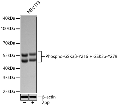
Western blot analysis of lysates from NIH/3T3 cells using Phospho-GSK3β-Y216 + GSK3α-Y279 Rabbit mAb at 1:1000 dilution incubated overnight at 4℃. NIH/3T3 cells were treated by λ-PP mixed solution (1μL) at 30℃ for 30 minutes. | Secondary antibody: HRP-conjugated Goat anti-Rabbit IgG (H+L) at 1:10000 dilution.|Lysates/proteins: 30 μg per lane.| Blocking buffer: 3 % nonfat dry milk in TBST.| Detection: ECL Basic Kit.| Exposure time: 20s.
Clonality:
Monoclonal
Host Species:
Rabbit
Tested Applications:
ELISA, WB
Conjugate:
Unconjugated
Species Reactivity:
Mouse
List Size:
100 uL