Affinity purification

Product Type
Research Area
Clonality
Host Species
Species Reactivity
Tested Applications
Conjugate
Product Badges
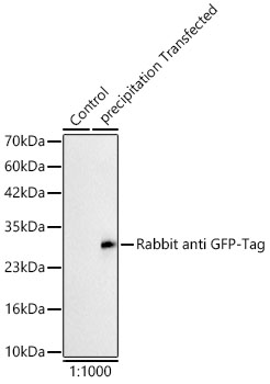
Western blot analysis of insect expressed GFP protein using HRP-conjugated Rabbit anti GFP-Tag at 1:1000 dilution incubated overnight at 4℃.|Lysates/proteins: 25 μg per lane.|Blocking buffer: 3% nonfat dry milk in TBST.|Detection: ECL Basic Kit|.Exposure time: 90s.
Clonality:
Monoclonal
Host Species:
Rabbit
Tested Applications:
ELISA, WB
Conjugate:
Unconjugated
Species Reactivity:
Species independent
List Size:
100 uL
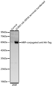
Western blot analysis of lysates from wild type (WT) and 293F cells transfected with SCRN1-HA-GFP(N terminal) using HRP-conjugated Rabbit anti HA-Tag mAb at 1:10000 dilution. |Lysates/proteins: 30 μg per lane.|Blocking buffer: 3% nonfat dry milk in TBST.|Detection: ECL Basic Kit.|Exposure time: 5s.
Clonality:
Monoclonal
Host Species:
Rabbit
Tested Applications:
WB
Conjugate:
Unconjugated
Species Reactivity:
Species independent
List Size:
100 uL
Western blot analysis of extracts of normal 293T cells, 293T transfected with EPCAM-His and 293T transfected with NPPA-His Protein, using HRP-conjugated Rabbit anti His-Tag mAb at 1:10000 dilution.|Lysates/proteins: 25μg per lane.|Blocking buffer: 3% nonfat dry milk in TBST.|Detection: ECL Basic Kit.|Exposure time: 0.09s.
Clonality:
Monoclonal
Host Species:
Rabbit
Tested Applications:
ELISA, WB
Conjugate:
Unconjugated
Species Reactivity:
Species independent
List Size:
100 uL
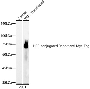
Western blot analysis of lysates from wild type (WT) and 293T cells transfected with YAP1 using HRP-conjugated Rabbit anti Myc-Tag mAb at 1:6000 dilution incubated overnight at 4℃.|Lysates/proteins: 25 μg per lane.|Blocking buffer: 3% nonfat dry milk in TBST.|Detection: ECL Basic Kit.|Exposure time: 1s.
Clonality:
Monoclonal
Host Species:
Rabbit
Tested Applications:
ELISA, WB
Conjugate:
Unconjugated
Species Reactivity:
Species independent
List Size:
100 uL
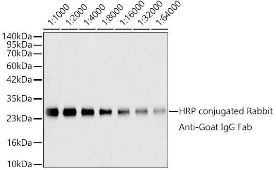
Western blot analysis of recombinant  goat IgG Fab protein using HRP-conjugated Rabbit anti-Goat IgG Fab mAb at 1:500-1:64000 dilution. Blot was incubated with the HRP conjugated antibody in the Blocking buffer (3% non-fat dry milk in TBST) at room temperature for 1 hour before detection using Enhanced chemiluminescence. |Lysates/proteins: Lane 1: molecular weight marker; Lanes 2-9: 100ng goat IgG Fab recombinant Protein per lane. |Detection: ECL Basic Kit. |Exposure time: 90s
Clonality:
Monoclonal
Host Species:
Rabbit
Tested Applications:
ELISA, WB
Conjugate:
HRP
Species Reactivity:
Goat
List Size:
100 uL
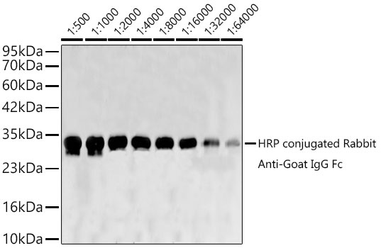
Western blot analysis of recombinant goat IgG Fc Protein using HRP-conjugated Rabbit anti-Goat IgG Fc mAb at 1:500-1:64000 dilution. Blot was incubated with HRP conjugated antibody in the Blocking buffer (3% nonfat dry milk in TBST) at room temperature for 1 hour before detection using Enhanced chemiluminescence. |Lysates/proteins: Lane 1: molecular weight marker; Lanes 2-9: 100ng goat IgG Fc recombinant Protein per lane. |Detection: ECL Basic Kit. |Exposure time: 15s.
Clonality:
Monoclonal
Host Species:
Rabbit
Tested Applications:
ELISA, WB
Conjugate:
HRP
Species Reactivity:
Goat
List Size:
100 uL
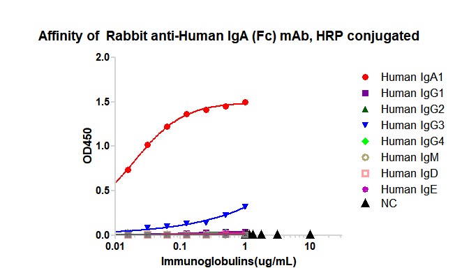
Dose response curve of HRP conjugated Rabbit anti-Human IgA (Fc) mAb measured by ELISA. 1 µg/mL of various immunoglobulins were coated to 384-well plate., blank wells without protein were used as negative control (NC). The coated plate was blocked and subsequently incubated with 25 µL of HRP conjugated Rabbit anti-Human IgA (Fc) mAb in a 2 fold serial dilution from 1 μg/mL to 7.8*10^-3 pg/mL, incubation was performed at room temperature for 1 hour. The ELISA result demonstrated that Rabbit Rabbit Anti-Human IgG4 has highly specific recognition of Human IgA (Fc) while no or minimal cross reactivity to Human IgG1、Human IgG2、Human IgG3、Human IgG4、Human IgM、Human IgE、Human IgD.
Clonality:
Monoclonal
Host Species:
Rabbit
Tested Applications:
ELISA
Conjugate:
HRP
Species Reactivity:
Human
List Size:
100 uL
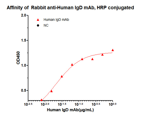
Dose response curve of HRP conjugated Rabbit Anti-Human IgD mAb measured by ELISA. 25 ng of Human IgD recombinant protein was coated in 384-well plate, blank wells without protein were used as negative control (NC). The coated plate was blocked and subsequently incubated with 25 µL of HRP conjugated Rabbit Anti-Human IgD mAb in a 2 fold serial dilution from 1 μg/mL to 7.8 pg/mL, incubation was performed at room temperature for 1 hour. EC50 value ( 2.10×10^-2 μg/mL) were calculated using 4 parameter logistic regression.
Clonality:
Monoclonal
Host Species:
Rabbit
Tested Applications:
ELISA
Conjugate:
HRP
Species Reactivity:
Human
List Size:
100 uL
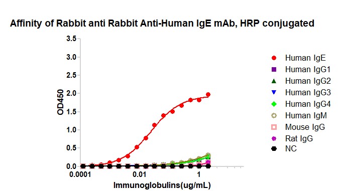
Dose response curve of HRP conjugated Rabbit Anti-Human IgE (Fc) mAb measured by ELISA. 1 µg/mL of various immunoglobulins were coated to 384-well plate., blank wells without protein were used as negative control (NC). The coated plate was blocked and subsequently incubated with 25 µL of HRP conjugated Rabbit Anti-Human IgE (Fc) mAb in a 2 fold serial dilution from 2 μg/mL to 6.1×10^-5 pg/mL, incubation was performed at room temperature for 1 hour. The ELISA result demonstrated that Rabbit Anti-Human IgE (Fc) mAb has highly specific recognition of Human IgE while no or minimal cross reactivity to Human IgG1、Human IgG2、Human IgG3、Human IgG4、Human IgM、Mouse IgG、Rat IgG.
Clonality:
Monoclonal
Host Species:
Rabbit
Tested Applications:
ELISA
Conjugate:
HRP
Species Reactivity:
Human
List Size:
100 uL
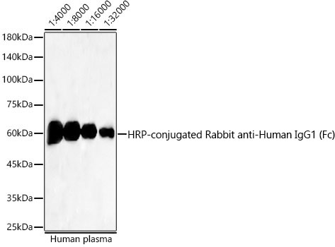
Western blot analysis of vlysates from Human plasma using HRP-conjugated Rabbit anti-Human IgG1 (Fc) mAb at 1:4000-1:32000 dilution.|Lysates/proteins: 25 μg per lane.|Blocking buffer: 3% nonfat dry milk in TBST.|Detection: ECL Basic Kit.|Exposure time: 30 s.
Clonality:
Monoclonal
Host Species:
Rabbit
Tested Applications:
ELISA, WB
Conjugate:
HRP
Species Reactivity:
Human
List Size:
100 uL